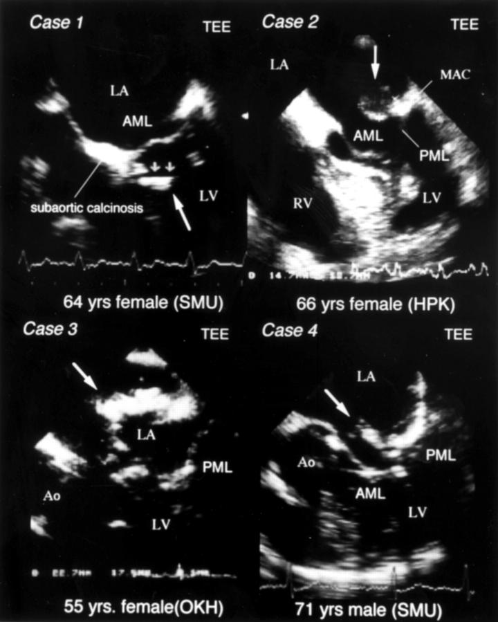
Figure 1 .
Case 1: transoesophageal echocardiography (TEE) showing a mobile intracardiac tumour (6 × 14 mm) with homogeneous hyperechoic characteristics, attached to the membranous portion of interventricular septum with tumour. Case 2: echocardiography after cerebral thromboembolism, showing a heterogenic high echoic mobile intracardiac tumour at the base of the posterior mitral leaflet (PML), and the rapid growth of the tumour (14.7 × 13.7 mm). Case 3: echocardiography showing rapidly growing intracardiac mass (14 × 12 mm) on posterior wall of left atrium (LA; mid-panel). Case 4: transoesophageal echocardiography showing a homogeneous high echoic mobile intra-atrial tumour located in anterior mitral valve (AMV) (8 × 12 mm). AML, anterior mitral leaflet; Ao, aorta; LV, left ventricle; MAC, mitral annular calcinosis; RV, right ventricle.

