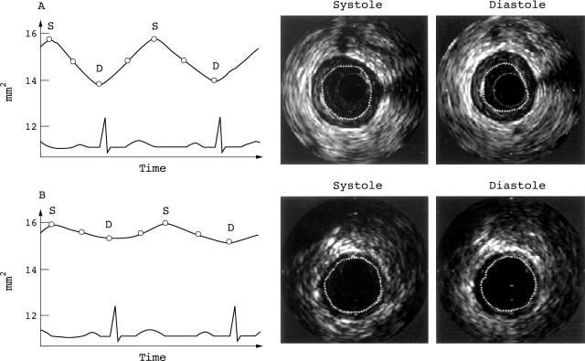
Figure 1 .
Comparison of the pulsatile variation of the cross sectional lumen area of a 77 year old male patient with an unstable lesion in his proximal left anterior descending artery (A) and of an age and target vessel matched patient with stable angina (B). The cyclic variation is graphically illustrated in the left panels, with the corresponding intravascular ultrasound images on the right. The change in luminal area over the cardiac cycle is more pronounced in the unstable patient. Empty circles indicate points of measurements. D, diastole; S, systole.

