
Fig. 4.
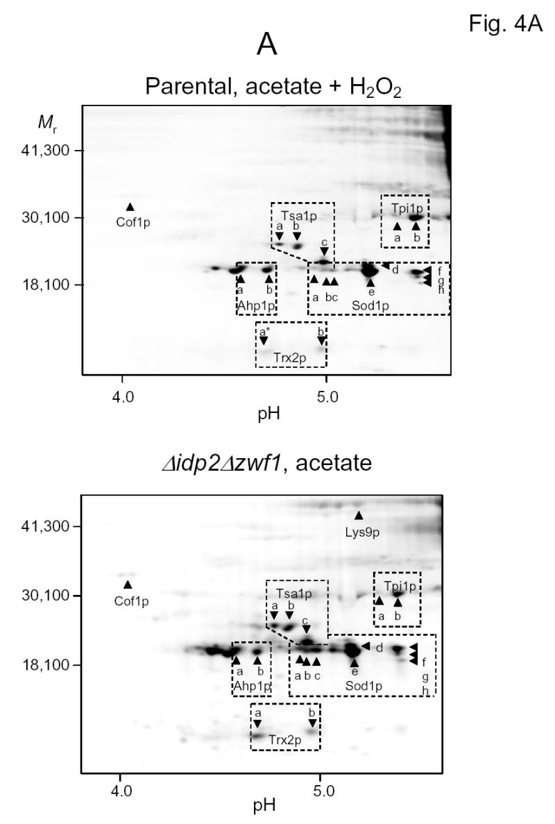
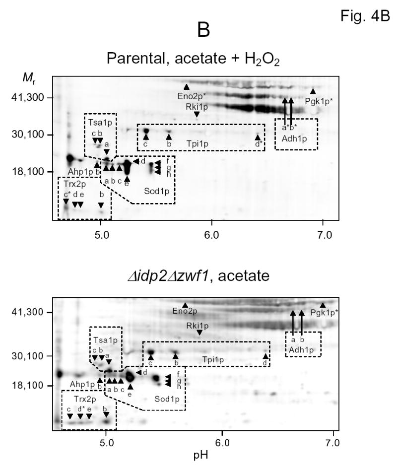
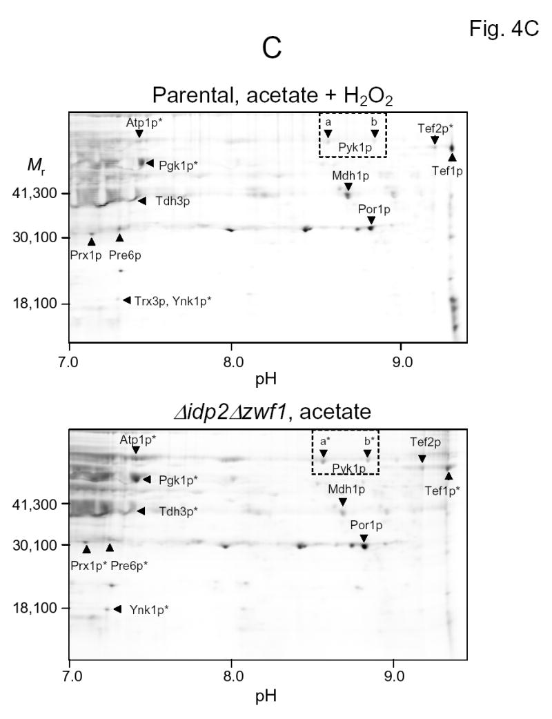
Two-dimensional gel electrophoresis of 6-IAF-labeled proteins from the parental strain shifted to acetate medium in the presence of hydrogen peroxide (top panels) and from the Δidp2Δzwf1 mutant strain shifted to acetate medium (bottom panels). Isoelectric focusing was conducted in the pH 3–6 range (A), in the pH 5–8 range (B), or in the pH 7–10 range (C). The indicated proteins were identified by mass spectrometry as described in Experimental Procedures. An asterisk indicates that labeling of a spot with 6-IAF was not above background in a sample.