
Figure 1.
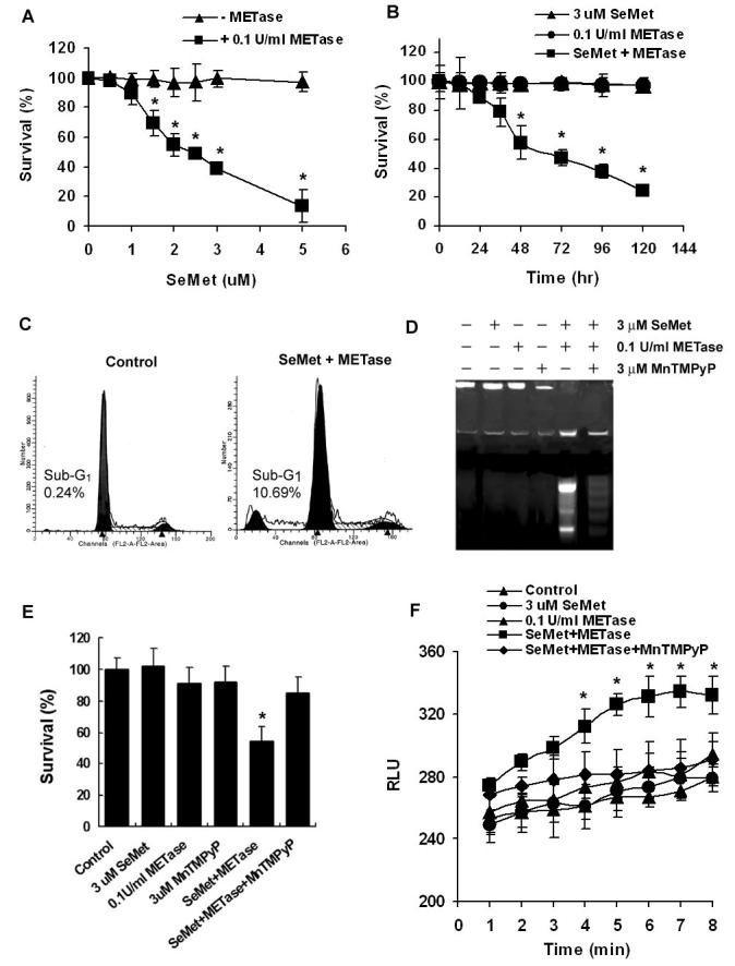
Effects of SeMet and METase on apoptosis and superoxide production in LNCaP cells. A, MTT assay demonstrating a dose-dependent effect of SeMet and METase on cell viability. Cells were treated with SeMet and METase for 5 days. B, MTT assay showing a time-dependent effect of SeMet, METase, or combination on cell viability. C, Flow cytometric analysis demonstrating apoptosis (sub-G1 cell population) induced by SeMet and METase. Cells were treated with 3 μM SeMet and 0.1 U/ml METase for 24 hr. D, Agarose gel electrophoretic detection of DNA fragmentation as a marker of cell apoptosis induced by SeMet and METase. Cells were treated with 3 μM SeMet, 0.1 U/ml METase, and 3 μM MnTMPyP alone or in combinations for 24 hr. E, Protection by MnTMPyP against cytotoxicity of SeMet and METase. Cells were treated with 3.0 μM SeMet, 0.1U/ml METase, 3.0 μM MnTMPyP alone or in combinations for 5 days and cell survival was measured by the MTT assay. F, Superoxide production in cells co-treated with SeMet and METase. Cells were treated with 3.0 μM SeMet and 0.1U/ml METase with/without 3.0 μM MnTMPyP and superoxide was measured using a chemiluminescence assay. RLU, relative light units. Data are presented as means ± SD of three independent experiments. * p<0.05 compared to no METase (A), SeMet or METase alone (B), and control or SeMet or METase (E and F).