
FIGURE 2. Stereo view of the ensemble of the 10 lowest energy structures for AbhN from B. subtilis and overlay of AbhN and AbrBN.
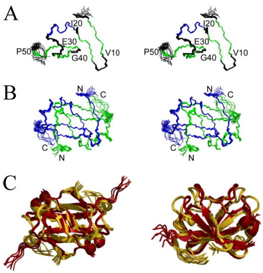
A, AbhN monomer colored by secondary structure; strands are colored green, whereas the helix is shown in blue. B, AbhN dimer colored by monomer. The superimposition is over residues 1–54 of each monomer, resulting in an r.m.s. deviation of 0.19 Å for backbone atoms. C, ribbon structure overlay of AbhN (gold ) and AbrBN (red ), highlighting the similarities and differences in the structures. Images were generated with Pymol (62).