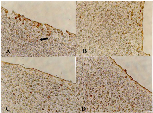
Figure 3.
Immunocytochemical detection of proliferating SMCs in neointima (×400). A: Control; B: Probucol; C: LXS; D: HXS. There are more PCNA-positive cells (→) in neointima of injuried coronary artery in control, indicating a state of cell proliferation, than those in different treatment groups, especially in HXS.

