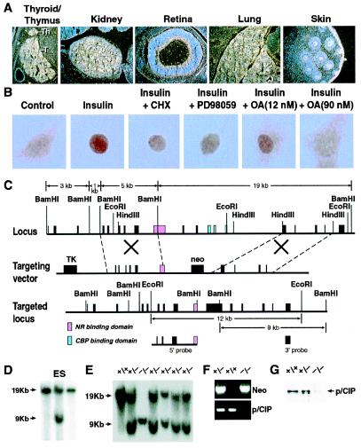
Figure 1.
Generation of p/CIP gene-deleted mice. (A) In situ hybridization analysis of p/CIP transcript in E15.5 embryo. Strong expression is detected in thyroid gland, thymus, kidney, retina, lung, and epidermis. (B) Cytoplasmic/nuclear localization of p/CIP. Under serum-free conditions, in Rat-1 cells, the immunoreactive p/CIP is largely cytoplasmic (control); addition of insulin (10−7 M) for 30 min causes most of p/CIP relocate to nucleus; pretreatment by cycloheximide (CHX; 10 μM) for 1 h or the mitogen-activated kinase kinase (MEK) inhibitor PD 18059 (10 μg/ml) and 12 nM okadaic acid did not alter the insulin effect; however, 90 nM okadaic acid reversed the insulin effect. (C) Strategy for homologous recombination of the p/CIP genomic locus. Probes of 5′ and 3′ are shown, and the regions targeted for deletion are nuclear receptor and CBP-binding domains. (D) Homologous recombination in ES cells. (E) Homologous recombination and proof of generation of gene-deleted mice. (F) PCR diagnostic strategy that is used to screen for genomic deletion. The primers used are from the deleted nuclear receptor binding domain. (G) Western blot analysis of kidney extract, showing the absence of p/CIP immunoreactive staining in p/CIP mice.

