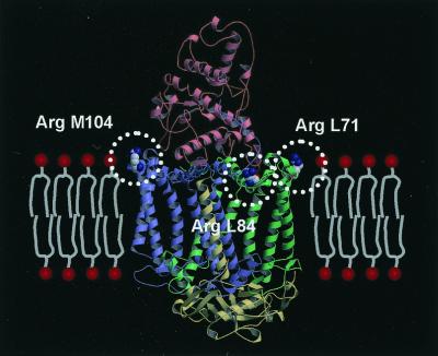
Figure 2.
Three Arg residues located at the membrane surface of the RC from Tch. tepidum. In the crystal structure, all three additional Arg residues (drawn by the space-filling model) are found to be located at the membrane surface. Each subunit is colored red (Cyt), green (L), blue (M), or yellow (H). The model of the membrane is placed on the ribbon model of the RC complex, and the head groups of the phospholipids are represented by the red circles. All three Arg residues are exposed to the solvent and located in a position from which they can interact with the head groups of the phospholipids.

