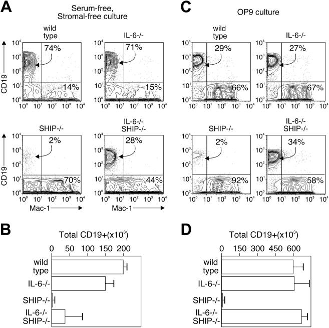
Figure 3.
Surface phenotype of single-cell cultures of progenitors. c-KithiSca-++ IL-7Rα– fraction containing uncommitted progenitors was isolated from wild-type, IL-6–/–, SHIP–/–, and IL-6–/– SHIP–/– animals. The sorted cells were cultured with serum-free, stromal cell-free condition (A-B) or with OP9 stromal cell (C-D). The results in panels C and D are the average and SD of 2 independent experiments, with each experiment having 3 individual wells. (A-B) The sorted cells were cultured with SCF, IL-7, and FL as previously described.22 The phenotype of colonies that emerged were examined by staining with anti-CD19 and anti–Mac-1 antibodies (A). Total CD19+ cells number after culture is shown in panel B. (C-D) The sorted cells were in the presence of OP9 stromal cells with SCF, IL-7, and FL for 12 days. The phenotype of colonies that emerged were examined by staining with anti-CD19 and anti–Mac-1 antibodies (C). Total CD19+ cell numbers after culture are shown in panel D. Numbers in panels A and C indicate the percentage of cells falling within the indicated gates. Bars in panels B and D represent the average and standard error of 4 replicate samples.

