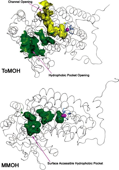
Figure 3.

Interior surface renderings of ToMOH (top) and MMOH (bottom) R-subunits. ToMOH channel van der Waals surface is shown in yellow; hydrophobic pockets in ToMOH and MMOH are shown in green. Protein backbones (Cα trace) are represented as gray ribbons, active-site iron atoms as magenta spheres, and side-chain ligands as blue sticks.
