
Fig. 5.
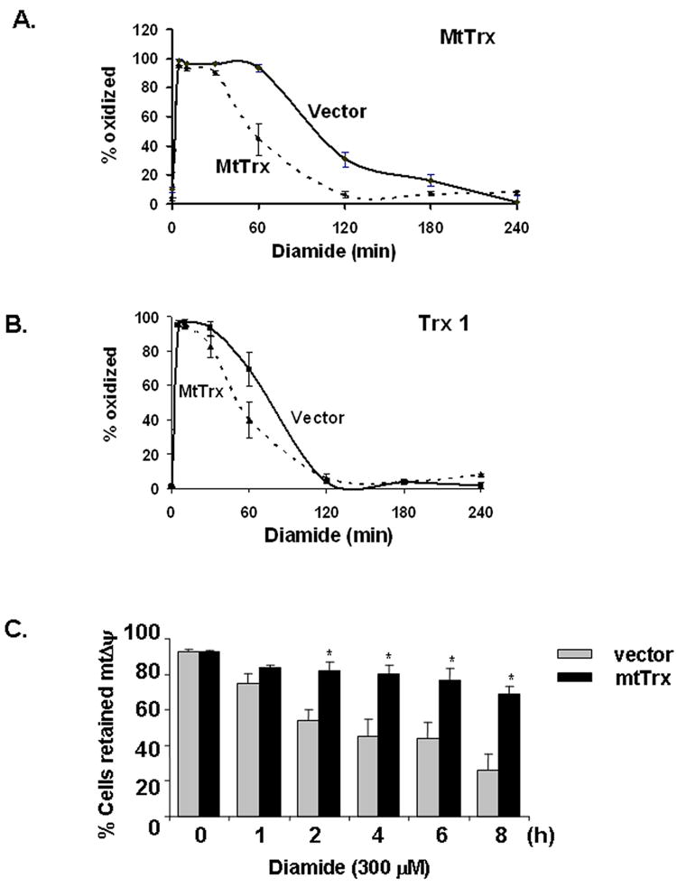
Overexpression of mtTrx in 143 B cells prevented oxidation of mtTrx and mitochondrial depolarization induced by diamide. 143 B cells transfected with empty vector or plasmid expressing mtTrx were treated with 300 μM diamide for indicated times. Cells were analyzed with Redox Western analysis to evaluate the redox status of mtTrx (A) and Trx 1 (B). The data were presented as the averages of five separate experiments (mean ± SEM). (C) Vector control and mtTrx-overexpressing cells were treated with 300 μM diamide for indicated time. MtΔψ was measured by TMRM staining followed by flow cytometry. The data presented are the average of five separate experiments (mean ± SEM). Significant differences from vector-transfected cells, determined by Student’s t-Test, are indicated as: *, P<0.05.