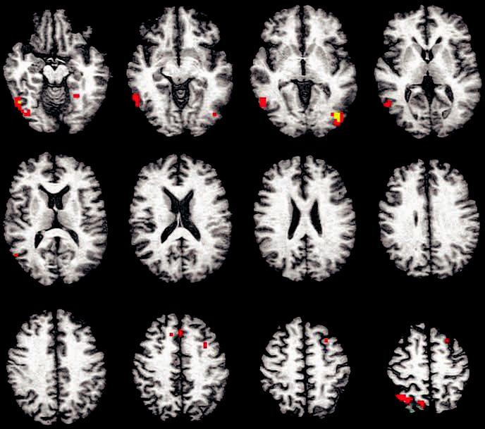
Figure 2.

Clusters of significant difference between APOE ε4 and ε3 participants for encoding-related brain response overlaid on a representative anatomic image in Talairach (1988) space (slices span from 12 inferior to 54 superior in 6 mm increments). Activations shown include voxels significant at p < 0.025 that are contained within a cluster of 13 or more voxels. Color scale represents effect sizes for the ε4 – ε3 difference in fit coefficient as measured by eta2 (signed to reflect the direction of the contrast) (red voxels: 0.50 < η2 ≤ 0.75; yellow voxels: 0.76 < η2 < 1.0).
