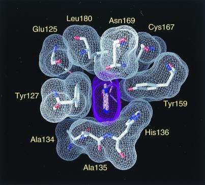
Figure 4.
The substrate-binding pocket of AAG. The ɛA base (purple surface) fits snugly into a pocket next to the enzyme active site. The base-binding pocket is viewed from the perspective of the protein, with the DNA helix oriented almost vertically behind the plane of the diagram. Met-149 and Cys-178 make additional van der Waals contacts to the ɛA base that are not shown.

