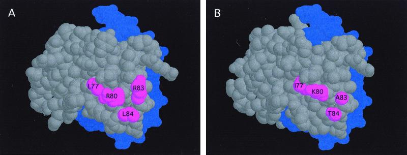
Figure 4.
Solvent-exposed surfaces of the C1 and P Myb domains. Modeling of the structure of the Myb domains of C1 (A) or P (B), based on the deduced structure of the R2R3 region of c-Myb (5). The DNA is shown in purple, and the four amino acids in C1 (L77, R80, R83, and L84) sufficient to transfer the interaction with R from C1 to P are shown in red. The position of G94 and R95 could not be precisely determined, although the polar nature of R95 makes it a candidate for a surface-exposed residue.

