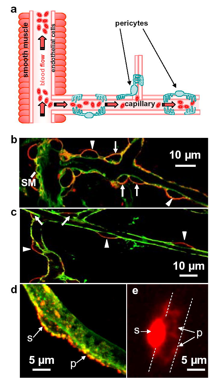
Figure 1.

Pericyte anatomy confers flow regulating capability downstream of arterioles. a Potential blood flow control sites in cerebral vasculature: arteriolar smooth muscle, and pericytes on capillaries. b Cerebellar molecular layer arteriole (left), surrounded by smooth muscle (SM), giving off a capillary. Capillary labelled with isolectin B4 (green); pericytes labelled for NG2 (red) are on the straight part of capillaries (arrow heads) and at junctions (arrows). c Retinal capillaries. d Soma (s) of cerebellar pericyte gives off processes (p) running along/around capillary. e Dye fill of retinal pericyte reveals processes running around capillary (dashed lines).
