
Figure 2.
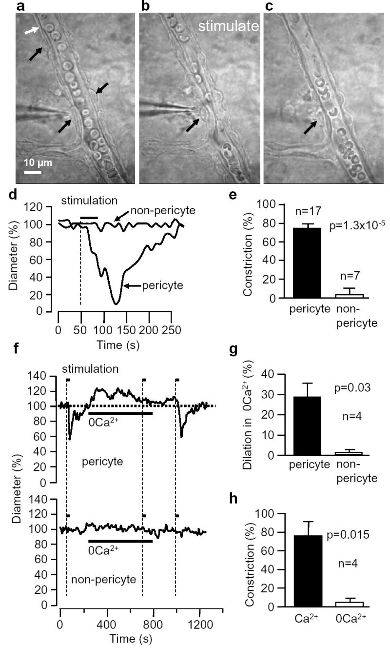
Electrical stimulation evokes a Ca2+-dependent localized constriction of retinal pericytes. a-c Capillary with pericytes (black arrows) before (a), during (b) and after (c) stimulation. Erthyrocytes are present within capillary; thin structures outside capillary are astrocyte endfeet. d Diameter of capillary in a-c at stimulated pericyte and a non-pericyte site (white arrow). e Mean (±s.e.m.) constriction when stimulating at pericyte or non-pericyte sites. f Effect of removing extracellular Ca2+ on resting diameter and response to pericyte stimulation, at pericyte and nearby non-pericyte sites. g Dilation produced by removing Ca2+. h Effect of Ca2+-removal on pericyte constriction, as in f.