
Figure 3.
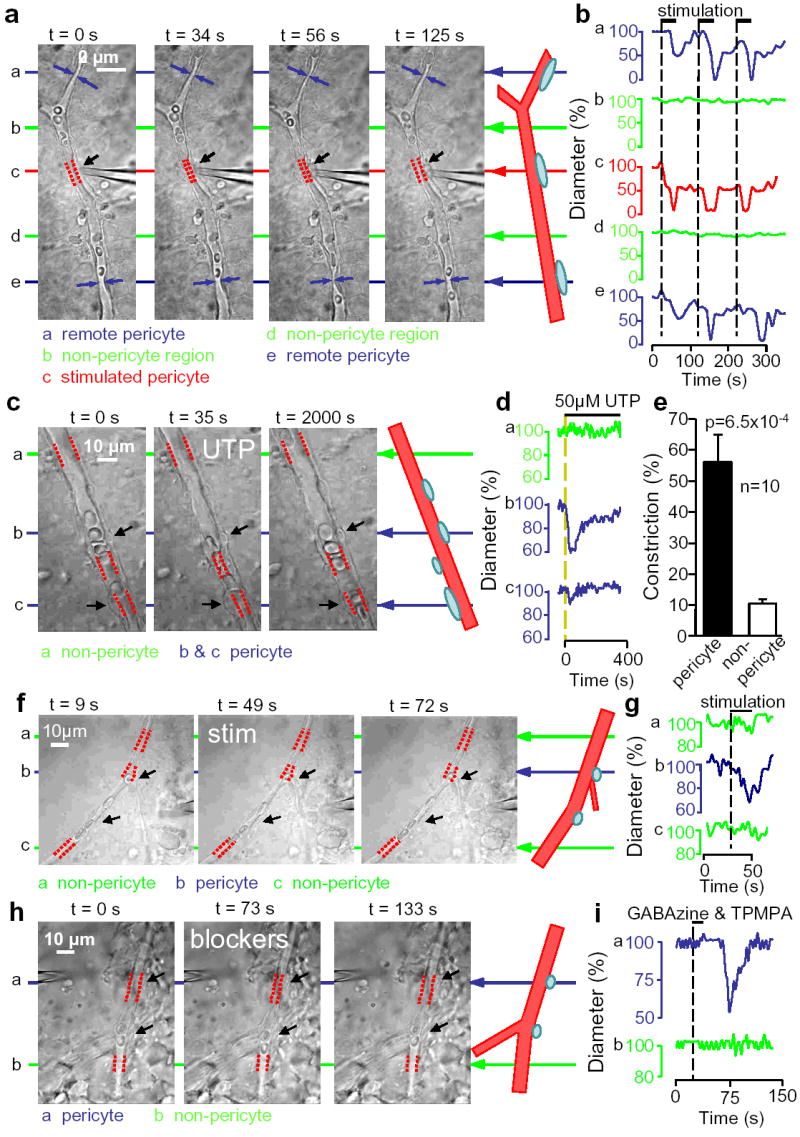
Propagation and transmitter-evocation of retinal pericyte constriction. a Electrically stimulating a pericyte (black arrow) evokes local constriction (red dashes show vessel diameter) followed by constriction of distant pericytes (blue). b No constriction of intervening non-pericyte regions (green). c,d UTP constricts two pericytes (arrows) but not at non-pericyte region. e Mean (±s.e.m.) UTP-evoked constriction. f,g Pericyte constriction (top arrows; lower arrows show another pericyte) evoked by electrical stimulation (electrode, right) near inner plexiform layer. h,i Constriction of pericyte (top arrows; lower arrows show another pericyte) evoked by puffing GABA receptor blockers (electrode, top left) near inner plexiform layer.