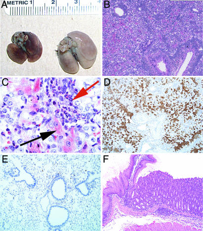
Fig. 1.
Pathology at 4 months after transfer of 3 × 10⁴ CD4 T cells into Rag2−/−mice. (A) Gross pathology of lungs from normal Rag2−/−mice and mice that had received 3 × 10⁴ CD4 T cells. (B and C) H&E stain (B) and Luna stain (C) for eosinophils (red arrow) and eosinophilic crystal-laden macrophages (black arrow). (D and E) Immunohistochemical anti-Ym1 stain of lungs from mice that had received 3 × 10⁴ CD4 T cells (D) and of lungs from normal mice (E). (F) H&E stain of junction of forestomach and glandular stomach from mice that had received 3 × 10⁴ CD4 T cells showing eosinophilic and lymphocytic infiltrate with parietal cell loss.

