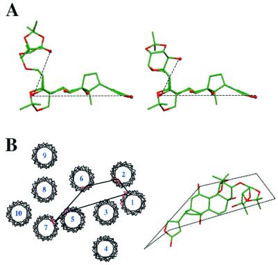
Figure 5.
Structural features of cardiac glycosides and the digitalis site. (A) Representatives of the two groups of closely related structures of OFDA at the digitalis site determined by 13C,19F-REDOR NMR. Carbon atoms are shown in green, oxygen atoms in red, and hydroxyl groups are represented as spheres for clarity. (B) The 10 putative transmembrane regions of the Na+/K+-ATPase α subunit were fit to the electron density map of Ca2+-ATPase (6), showing in red the mutation sites conferring ouabain resistance to HeLa cells. One possible structure of OFDA is shown alongside the protein model to illustrate the comparative dimensions of the cardiac glycosides and the surface of the α subunit, and a possible docking orientation.

