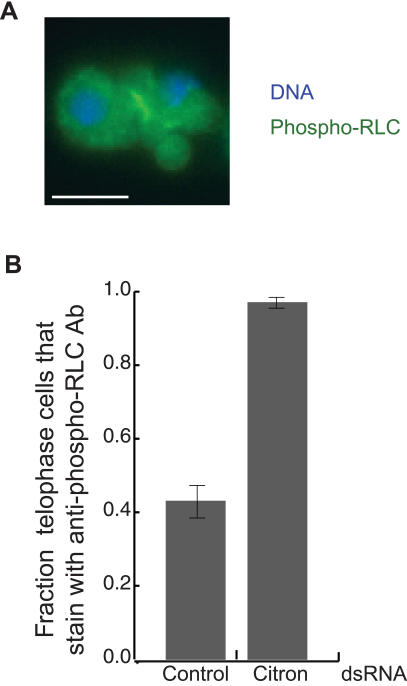
Figure 4. Citron is not essential for RLC phosphorylation.
(A) Immunofluorescent localization of DNΑ (blue) and phospho-RLC (green) in Citron depleted cells. Notice the membrane blebs in the telophase furrow as well as the anti-phospho-RLC antibody staining in the midbody. Scale bar = 5 µm. (B) Depletion of Citron probably affects the structure of telophase midbodies. S2 cells were depleted of Citron by treatment with RNAi for 5 days and then fixed and stained with an anti-phospho-RLC antibody. Late telophase cells were scored for antibody staining (mean, ±SE for 30 cells). While less than half of all control cells were stained, nearly all Citron depleted cells were stained.

