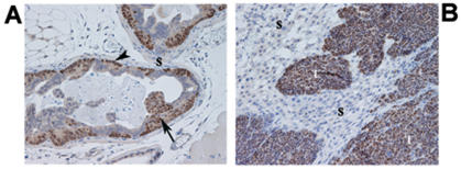
Figure 1. Histological appearance of PIN and invasive CR2-TAg prostate cancer lesions.
(A) prostate glands of a 10-week old CR2-TAg mouse showing flat and tufted patterns of PIN (arrowhead and arrow, respectively), and a paucicellular stroma (S); (B) invasive cancer lesion from a 24-week old mouse where PIN acini have been replaced by solid tumor (T) and an abundant cellular, reactive stroma (S) composed primarily of fibroblasts/myofibroblasts as assessed by vimentin/actin smooth muscle staining (data not shown).
Tissue sections were stained using anti-SV40 antibody (brown) and counterstained with haematoxylin.
Magnification 100×.

