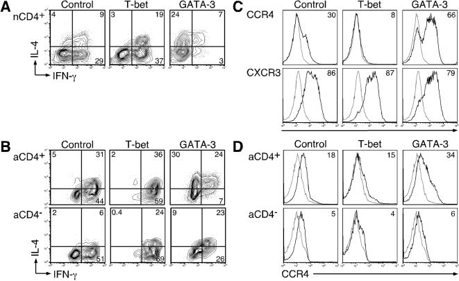
Figure 4. Functional profile of NKT cell subsets ectopically expressing T-bet or GATA-3.
NKT cells were activated under non-polarizing conditions as in Figure 2, transduced at the time of activation with either the control lentiviral vector or vectors expressing T-bet or GATA3, and expanded in IL-2-containing media.
One representative donor out of 3 is shown for each.
(A) Intracellular stain of nCD4+ NKT cells was performed as in Figure 2 and cells were co-stained with anti-mCD24 (FITC) to identify transduced cells.
(B) Intracellular cytokine stains of aCD4+ or aCD4− NKT cells were performed as in 4A.
CD4 expression was determined by co-staining with anti-CD4 (biotin followed with strepavidin PerCP-Cy 5.5).
(C) CCR4 or CXCR3 expression on T-bet- or GATA-3-transduced nNKT cells was assayed as in Figures 3A and 3B, and cells were co-stained with anti-mCD24 (FITC) as a marker for transduction.
(D) CCR4 expression of T-bet or GATA-3-transduced aCD4+ or aCD4− NKT cell was examined as in 4C, and cells were stained with anti-CD4 (PE) to differentiate between CD4+ and CD4− NKT cells.

