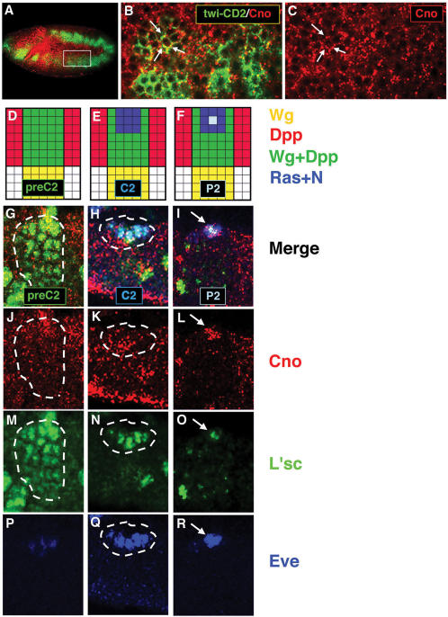
Figure 1.
Cno is expressed in the mesoderm throughout progenitor specification.
(A) Confocal immunofluorescence showing a lateral view of a late stage 10 embryo.
Cno (red) is detected in the mesoderm (green).
(B, C) Higher magnification of two hemisegments (63×; inset in A) reveals punctuate Cno expression at submembrane locations (arrows).
(D–F) Diagrams show the most dorsal part of one hemisegment and the signals involved throughout dorsal progenitor specification.
(G–R) Confocal immunofluorescences showing high magnification (63×) of the most dorsal part of one hemisegment.
(D, G, J, M) Cno is expressed in the dorsal mesodermal region pre-patterned by Wg and Dpp (preC2) along with L'sc.
(E, H, K, N, Q) Cno is detected in the equivalence group in which Ras is locally activated (C2) restricting L'sc to this cluster and activating Eve.
(F, I, L, O, R) Cno is expressed in the progenitor (P2) singled out from this cluster after N-promoted lateral inhibition.

