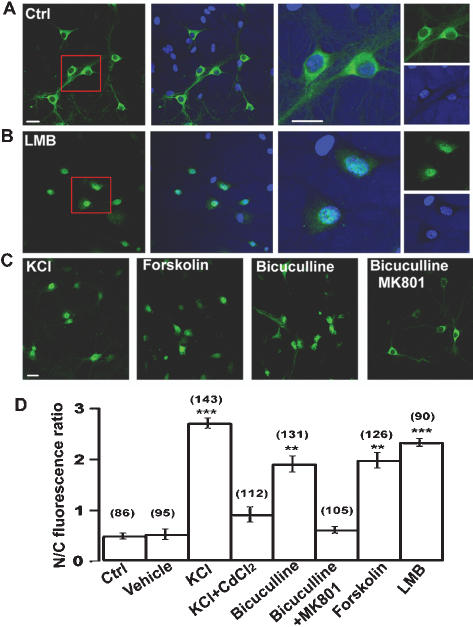
Figure 2. Activity-dependent TORC1 nuclear translocation in cultured hippocampal neurons.
(A) Representative immunocytochemistry staining of TORC1 under control condition with lower magnification (left panels).
Hochest was used for nuclear staining.
Scale bar: 20 µm.
High magnification of TORC1 staining and Hochest for nuclear staining (right panels).
Scale bar: 20 µm.
(B) Representative immunocytochemistry staining of TORC1 after LMB treatment at lower magnification (left panels) and high magnification (right panels).
(C) Representative immunocytochemistry staining of TORC1 after treated with KCl, forskolin, bicuculline and bicuculline plus MK801.
(D) Statistical analysis of TORC1 distribution after the indicated treatments.
Error bars indicate SEM, data in each group were obtained from four independent experiments.
The number associated with each column refers to the number of neuron analysed in each treatment.
**, p < 0.01; ***, p < 0.001, compared to control group.

