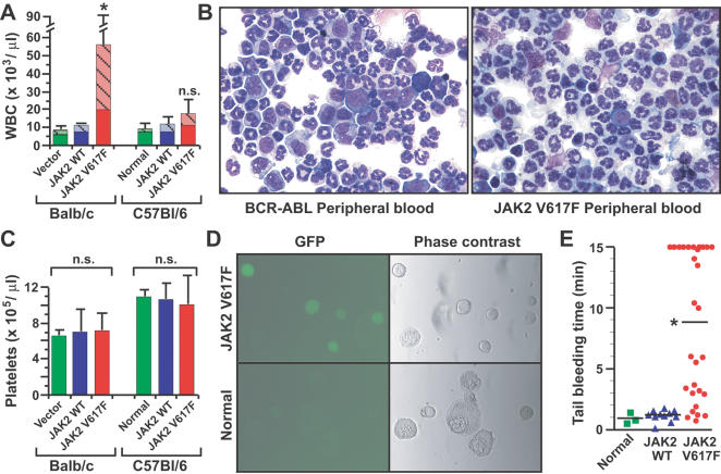
Figure 2. Effect of JAK2 V617F on leukocyte and platelet counts.
(A) Peripheral blood leukocyte counts for the three cohorts in Balb/c and B6 backgrounds as in Figure 1.
The difference in leukocyte counts between JAK2 V617F and JAK2 WT recipients was significant for the Balb/c cohort (P = 0.0039) but not for B6 (P = 0.0542, unpaired t-test).
The hatched portion of the histogram represents the percentage of the total leukocyte count comprised by neutrophils.
(B) Wright-Giemsa-stained cytospins of peripheral blood leukocytes from representative mice with BCR-ABL-induced CML-like MPD (left) or JAK2 V617F-induced PV-like MPD (right).
Note the predominance of mature neutrophils and lack of immature myeloid elements (myelocytes and promyelocytes) in the JAK2 V617F recipient.
(C) Platelet counts for the groups in (A).
There was no significant difference in platelet counts between the three cohorts in either stain.
(D) GFP fluorescence (left panels) and phase contrast images (right panels) of purified megakaryocytes from recipients of JAK2 V617F-transduced BM (top panels) or normal control mice (bottom panels).
Note the GFP fluorescence in the majority of megakaryocytes from JAK2 V617F recipients.
(E) Tail bleeding time (Balb/c cohort) for the three groups.
The mean bleeding time (bar) of JAK2 V617F recipients was significantly longer than that of vector or JAK2 WT recipients (P = 0.0328 and P = 0.0002, respectively, unpaired t-test).

