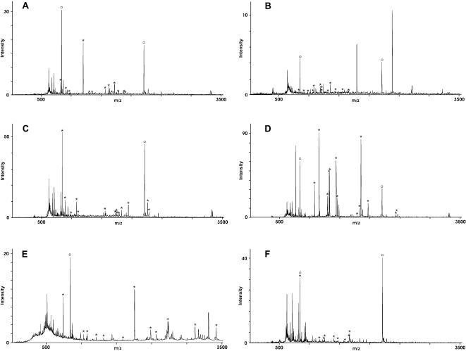
Figure 4. Identification of L. donovani antigens by mass spectrometry.
The protein spots in the silver-stained gel that had been assigned to antigen spots in the corresponding Western blot were excised, destained and incubated with trypsin.
The resulting fragments were extracted from the gel pieces and analyzed by MALDI-TOF-MS.
Panels A–F show the peptide mass fingerprints (PMF) of the proteins in spots 1–6, respectively.
Upon processing via MASCOT, the antigens were identified as HSP70 (spots 1, panel A and spot 2, panel B), gp63 (spot 3, panel A), EIF-4a (spot 4, panel C), Ef2 (spot 5, panel D) and grp78 (spot 6, panel E).
The fragment masses that could be matched to theoretical trypsin digests of the identified proteins are indicated by asterisks.
Open circles indicate the autolytic fragments of trypsin that were used for internal calibration.

