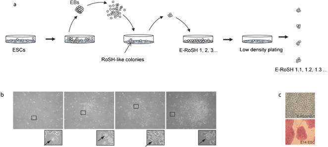
Figure 1. Derivation of E-RoSH cell lines.
(a) ESCs were plated singly on methycellulose based media to form EBs.
At day 3–6, EBs were harvested, dissociated by collagenase and cultured as a monolayer on gelatinized feeder plate.
RoSH-like colonies with adherent fibroblast-like cells and ring-like structures were selected and propagated on gelatinized plates to generate E-RoSH 1, 2, 3… Each of the cultures were then plated at a low density of 10–100 cells per 10 cm plate and single RoSH like colonies were picked to established sublines, E-RoSH 2.1, 2.2, 2.3. .. etc;
b) A putative RoSH-like colony consisting of adherent short fibroblast-like cells with characteristic ring-like cells (inset) expanding over time;
c) Alkaline phosphatase staining of E-RoSH2.1 and its parental E14 ES cells.

