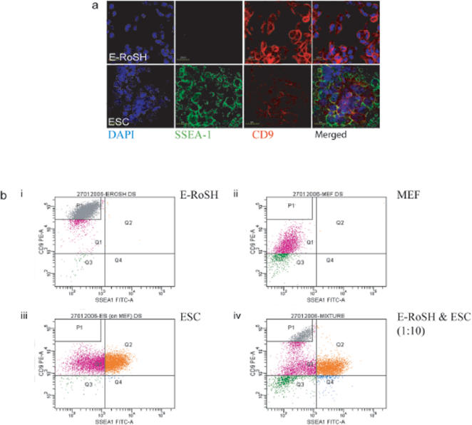
Figure 4. Identifying selectable surface antigens for the isolation of putative RoSH-like cells from differentiating ESCs.
a) Confocal microscopy of E-RoSH2.1 cells (top) and E14 ESCs (bottom).
The cells were counterstained with DAPI, a nuclear stain after immunostaining for anti-SSE4-1 antibody conjugated with FITC and anti-CD9 antibody conjugated with PE.
b) FACS analysis of i) E-RoSH2.1 cells, ii) E14 ESCs, iii) murine embryonic fibroblast (MEF) and iv) 10∶1 mixture of E14 ESCs and E-RoSH2.1 cells.
The cells were labelled with anti-CD9 antibody conjugated with PE and anti-SSE4-1 antibody conjugated with FITC, and analyzed on a FACS Aria using FACS Diva software (BD Biosciences Pharmingen, San Diego, CA).

