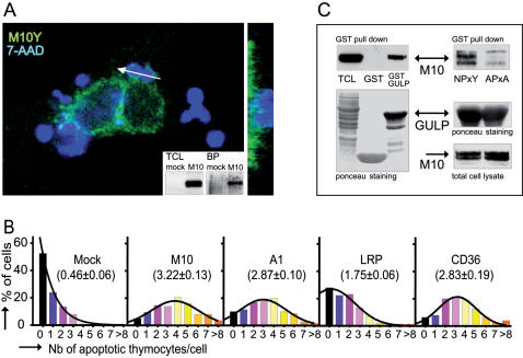
Figure 2. Functional assessment of MEGF10 as an engulfment receptor.
(A) MEGF10 is expressed at the cell surface and clusters around cell corpses during engulfment. Confocal optical X-Y sections show the localization of MEGF10 EYFP (M10Y - pseudo color green) in transfected HeLa cells challenged with 7-AAD labelled apoptotic thymocytes (pseudo color blue). The white arrow indicates the location of the X-Z plan shown on the right. The surface localization was confirmed by the analysis of surface biotinylation as shown in the insert. Western blot of total cell lysates probed with an anti-GFP antibody (TCL, left panel) are compared to blots of biotinylated proteins (BP, right panel). Mock transfected cells (mock), and MEGF10 EYFP transfected (M10) HeLa cells. (B) The expression of MEGF10 enhances the phagocytic ability of HeLa Cells. In vitro phagocytosis assays were carried out on HeLa cells transfected with the indicated engulfment receptors. Results are expressed as distribution histograms. Percentages of cells, scored on ≥100 transfected cells, are plotted against the number of tethered/ engulfed apoptotic thymocytes. The phagocytic index, computed out of at least 4 individual experiments, is indicated in brackets. Mock: mock transfected cells; M10: MEGF10; A1: ABCA1; LRP: LRP-1. (C) MEGF10 can interact with GULP as assessed by GST pull down. Left panel: lysates of HeLa cells transfected with MEGF10 EYFP were incubated with bacterially produced GST or GST-GULP fusion protein before fractionation on SDS-PAGE and blotting onto nitrocellulose membrane. Total proteins were visualized by Ponceau S staining (lower panel) whereas MEGF10 binding to GULP was detected by hybridization with an anti-GFP antibody (upper panel –TCL: Total cell lysate). Right panel: GULP and MEGF10 interact via the NPxY motif since its mutation to APxA abrogates binding (upper panel - NPxY: MEGF10 EYFP-NPxY, APxA: MEGF10 EYFP-APxA). Equivalent amounts of bacterially produced GST-GULP (middle panel) were incubated with equivalent amounts of MEGF10 expressed in transfected cells (lower panel). MEGF10 detection was performed by probing with an anti-GFP antibody on total cell lysate (lower panel) or on pulled down samples (upper panel).

