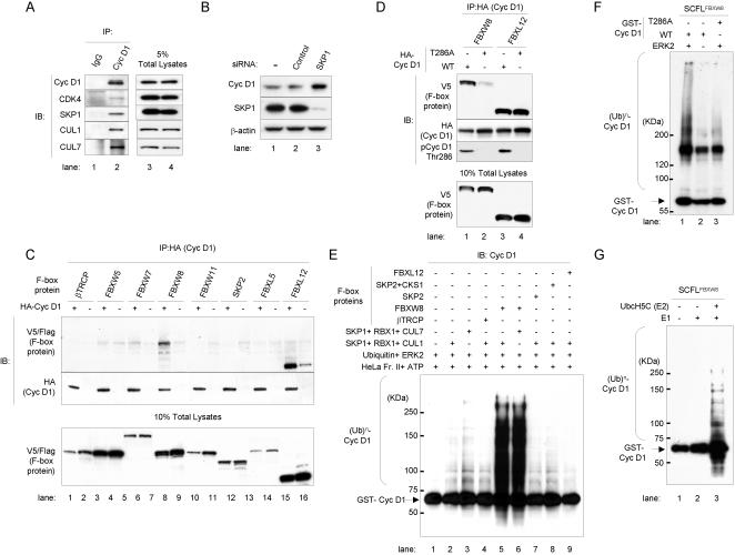
Figure 3.
FBXW8 ubiquitinates cyclin D1 in a Thr286 phosphorylation-dependent manner. (A) IP-IB analysis (left). Protein from exponentially growing HCT 116 cells was precipitated with antibodies to cyclin D1 or IgG. Immunoprecipitates were subjected to SDS-PAGE and sequentially blotted with cyclin D1, CDK4, SKP1, CUL1 and CUL7 antibodies. IB analysis with 5% of total cell lysates was provided as a control (right). (B) IB analysis following depletion of SKP1 expression for 48 hrs after treatment with SKP1 siRNA double-strand oligonucleotides in HCT 116 cells. Non-targeting siRNA (Control) and mock transfection (−) served as controls. (C) IP-IB analysis. Twenty-six F-box full-length encoding cDNAs were cloned into V5 or Flag epitope tag expression vectors. These V5 or Flag-tagged F-box protein DNA plasmids were transfected together with HA-tagged cyclin D1 (HA-Cyc D1) and CDK4 expression vectors into T98G cells. Cells were collected 24 hrs later. Samples were precipitated with a HA epitope tag antibody. Immunoprecipitates were subjected to SDS-PAGE and subsequently stained with V5 or Flag (F-box proteins), HA (Cyc D1) antibodies. IB analysis with 10% of total cell lysates was provided (bottom). (D) IP-IB analysis (top). V5-tagged F-box protein DNA plasmids were transiently transfected together with either HA-tagged cyclin D1 (Cyc D1) wild type (WT) or T286A mutant, and CDK4 expression vectors in T98G cells respectively. Samples were precipitated with a HA epitope-tag antibody. Immunoprecipitates were subjected to SDS-PAGE and subsequently blotted with V5 (F-box proteins), HA (Cyc D1), and Thr286 phosphorylated cyclin D1 (pCyc D1 Thr286) antibodies. IB analysis with 10% of total cell lysates was provided for comparison (bottom). (E) In vitro ubiquitination assay. In vitro translated F-box proteins with recombinant GST-full-length cyclin D1 (Cyc D1) wild type, HeLa cell extracts Fraction II with ATP, Ubiquitin and ERK2, and in vitro-translated either SKP1, RBX1 and CUL1, or SKP1, RBX1 and CUL7 proteins were incubated at 30°C for 2 hrs. Samples were separated by SDS-PAGE and immunoblotted with a cyclin D1 antibody. (F) In vitro polyubiquitination of cyclin D1 through the SCF-like (SCFL) complex FBXW8 (SKP1-CUL7-FBXW8-RBX1/SCFLFBXW8). WT or T286A GST- Cyc D1 was incubated in the presence of purified ERK2 (lanes 1, and 3) or its absence (lane 2) at 30°C for 2 hrs. Samples were separated by SDS-PAGE and immunoblotted with a cyclin D1 antibody. Asterisk indicates non-specific bands. (G) Reconstitution of polyubiquitination of cyclin D1 through SCFLFBXW8 in vitro using purified E1 and E2. GST-WT Cyc D1 was incubated with recombinant SCFLFBXW8 in the presence or absence of E1 and E2/UbcH5C. Samples were separated by SDS-PAGE and immunoblotted with a cyclin D1 antibody.

