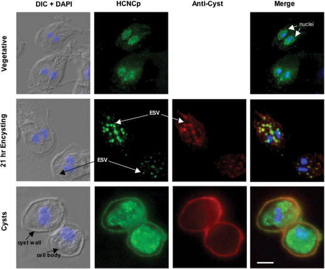
Figure 3. Traffic of HCNCp And Cyst Proteins During Growth And Differentiation.
Differential interference contrast (DIC) merged with DAPI images are shown to the left of each panel, HCNCp in green, anti-cyst proteins in red, and nucleic acid in blue (DAPI).
In trophozoites, HCNCp localized to nuclei and nuclear envelope/ER.
During encystation HCNCp co-localized with cyst proteins in encystation secretory vesicles (ESV) and to the cyst wall of water-resistant cysts.
In mature cysts, most of the HCNCp was within the cell body.
Scale bar is 5 µM.

