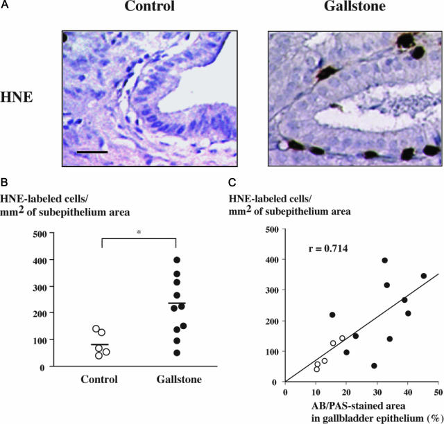
Figure 2.
Neutrophil infiltration of the gallbladder subepithelium in gallstone disease. A: Photomicrographs of gallbladder tissue sections labeled for HNE showing no labeling in a control specimen (left) and neutrophil infiltration in the subepithelium of a gallbladder with gallstones (right). Bar = 50 μm; original magnification, ×200. B: HNE-labeled cells were counted in the subepithelium of gallbladder specimens from controls (n = 5, open circles) and from individuals with gallstones (n = 10, solid circles). Horizontal bars represent median values; *P < 0.05. C: Correlation between the number of HNE-labeled cells in the subepithelium and AB/PAS-stained areas in the epithelium of gallbladders from controls (open circles) and from individuals with gallstones (solid circles); r = 0.714; P < 0.05.

