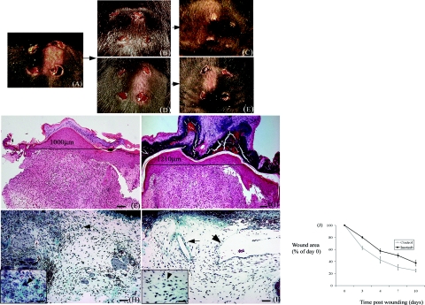
Figure 1.
Wound healing is impaired in mice treated with imatinib. A: Punch wounds (4 mm3) were made on the back of anesthetized mice. After 3 days, wound diameters are noticeably smaller in control animals (B) in comparison with imatinib-treated animals (D). After 7 days, wound diameters in control wounds (C) are clearly reduced compared with imatinib-treated wounds (E). Analysis of sections stained with H&E confirmed that after 7 days, distance between wound margins was greater in imatinib-treated animals (G) compared with control animals (F). Staining of sections with Masson’s trichrome confirmed that granulation tissue in treated animals was poorly formed and relatively hypocellular (I, arrows) compared with control animals (H, arrows). Granulation tissue in control animals was characterized by the formation of small blood vessels (H, inset, arrowhead), which were not present in imatinib-treated wounds (I, inset, arrowhead). Quantification of wound diameter throughout 10 days after injury. Results represent the mean ± SEM. Between 3 and 7 days after wounding, the difference in wound diameter was significant (P < 0.05). Scale bars = 100 μm (F, G); 50 μm (H, I); 10 μm (insets).

