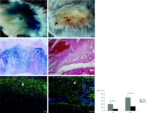
Figure 7.
Collagen type I gene promoter activity is reduced in imatinib-treated animals. Seven days after injury, wounds were excised from mice and stained with 1 mg/ml X-gal. Control wounds show uniformly intense X-gal staining (A, arrow), whereas staining in tissue from imatinib-treated mice was significantly weaker and confined to the wound margins (B, arrow). C: In histological sections, X-gal staining could be observed in fibroblastic cells throughout the granulation tissue (arrowheads). D: In sections from imatinib-treated animals, relatively fewer blue cells were present and were restricted to the wound margins (arrowhead). G: Imatinib-mediated reduction in collagen transgene activity was confirmed by quantification of transgene activity in whole wounds. Results represent the mean ± SD. *P < 0.01. Immunofluorescence staining of 14-day-old wounds confirming that expression of collagen protein was concordantly reduced in imatinib-treated wounds (F, arrow) relative to control tissue (E, arrow). Scale bar = 50 μm.

