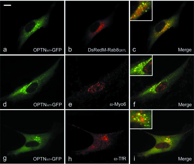
Figure 5.
Colocalization of the OPTN foci with other proteins. For colocalization with Rab8, TM cells were co-transfected with pOPTNWT-GFP (a; green) and pDsRedM-Rab8Q67L (b; red). For colocalization with myosin VI (e; red) and transferrin receptor (h; red), TM cells were immunostained with antibody to myosin VI (α-Myo6; e; red) and transferrin receptor (α-TfR; h; red) after transfection with pOPTNWT-GFP (d–i; GFP in green). Colocalization is observed in merged images (c, f, and i). The perinuclear region is shown at a higher magnification in insets. Images were taken using a 63× oil objective. Bar = 10 μm.

