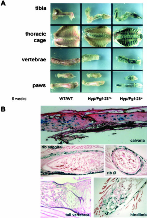
Figure 1.
Expression of Fgf-23 by β-galactosidase staining at 6 weeks. A: Shown are various skeletal elements including tibia, thoracic cage, vertebrae, and paws of wild-type (WT/WT) mouse, an Fgf-23 heterozygous mouse in a Hyp mouse background (Hyp+/−/Fgf-23+/−), and an Fgf-23 homozygous mutant mouse in a Hyp mouse background (Hyp+/−/Fgf-23−/−). Please note the difference in intensity of β-galactosidase staining in Hyp+/−/Fgf-23+/− versus Hyp+/−/Fgf-23−/− bones. B: Represented are frozen sections of stained Hyp+/−/Fgf-23−/− bones such as calvaria, ribs (saggital and transverse), tail vertebrae, and tibia/hindlimb; specific blue staining is only evident in osteocytes of intramembranous and endochondral formed bones. No staining is evident in osteoblasts or cartilaginous areas.

