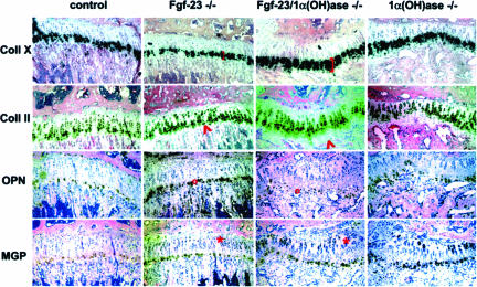
Figure 5.
In situ hybridization with riboprobes for collagen type X (Coll X), collagen type II (Coll II), osteopontin (OPN), and matrix gla protein (MGP) on sections from tibia of control, Fgf-23−/−, Fgf-23−/−/1α(OH)ase−/−, and 1α(OH)ase−/− at 6 weeks. Brackets depict the size of the zone of hypertrophic chondrocytes. Red arrowheads point to an area of hypertrophic chondrocytes that is only present in Fgf-23−/−/1α(OH)ase−/− and 1α(OH)ase−/− bones. Red circles show the decrease in OPN expression in the ricketic phenotype, and red asterisk depicts an expansion of MGP expression in the growth plate of Fgf-23−/−/1α(OH)ase−/− and 1α(OH)ase−/− bones.

