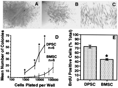
Figure 1.
Colony-forming efficiency and cell proliferation in vitro. Representative high (A) and low (B) density colonies after 14 days. The morphology is typical of fibroblast-like cells (C). The incidence of colony-forming cells from dental pulp tissue and bone marrow at various plating densities indicates that there are more clonogenic cells in dental pulp than in bone marrow (D). The number of BrdUrd-positive cells were expressed as a percentage of the total number of cells counted for DPSCs and BMSCs (E). Statistical significance (*) was determined by using the Student's t test (P ≤ 0.05).

