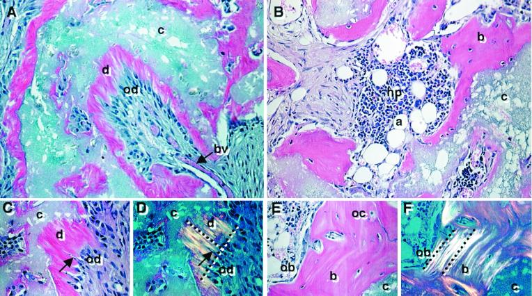
Figure 4.
Developmental potential in vivo. Cross sections are representative of DPSC transplants (A, C, and D) and BMSC transplants, (B, E, and F) 6 weeks posttransplantation and stained with hematoxylin and eosin. In the DPSC transplants, the HA/TCP carrier surfaces (c) are lined with a dentin-like matrix (d), surrounding a pulp-like tissue with blood vessels (bv) and an interface layer of odontoblast-like cells (od) (A). A magnified view of the dentin matrix (d) highlights the odontoblast-like layer (od) and odontoblast processes (arrow) (C). Polarized light demonstrates perpendicular alignment (dashed lines) of the collagen fibers to the forming surface (D). In BMSC transplants, lamellar bone (b) is formed on the HA/TCP surfaces (c) and surrounds a vascular, hematopoietic marrow organ (hp) with accumulated adipocytes (a) (B). A magnified view shows that the new bone contains osteocytes (oc), embedded within the calcified matrix, and osteoblasts (ob) lining the bone surfaces (E). With polarized light, collagen fibrils are seen to be deposited parallel with the forming surface (dashed lines) (F).

