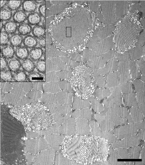
Figure 2.
Cav-2-deficient skeletal muscle fibers display tubular aggregate formation. Low-magnification electron micrograph of skeletal muscle from 3-month-old Cav-2-deficient mice. Note the multiple examples of tubular aggregate formation. A high-magnification inset reveals the crystalline-like organization of tubular aggregates. Scale bar for low-magnification image = 1 μm; scale bar in insert = 50 nm.

