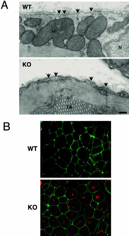
Figure 4.
Cav-2-deficient skeletal muscle fibers retain the ability to form caveolae and exhibit normal Cav-3 levels and distribution. A: Electron micrographs demonstrate that both WT and Cav-2 KO skeletal muscle have abundant caveolae (arrows) at the plasma membrane. Note the presence of a tubular aggregate (TA) in the Cav-2 KO micrograph. M, mitochondria. N, nucleus. Scale bar in KO = 250 nm and applies to both images. B: Cav-3 (green) is localized at the plasma membrane in WT and Cav-2-deficient mice skeletal muscle. Staining with an antibody to SERCA-1 (red) reveals extensive tubular aggregate formation in Cav-2 deficient but not in WT skeletal muscle fibers.

