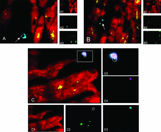
Figure 2.
Confocal microscopic examination of the unaffected myocardium of an AMI patient (two-dimensional reconstruction with pseudocolors). A: Triple immunostain for cardiomyocytes (sarcomeric actin, Texas Red; A1), macrophages (CD 68, cumarin; A2), and CP OMP-2 antigens (FITC; A3). Cardiomyocytes observed gave rise a strong positive reaction for CP OMP-2 in the cytoplasm (double positivity, appears as yellow stain), whereas the rare macrophages scattered among cardiomyocytes are negative for CP antigen (arrow). B: Triple immunostain for cardiomyocytes (sarcomeric actin, Texas Red; B1), T lymphocytes (CD3, cumarin; B2), and CP OMP-2 antigens (FITC; B3). Two-dimensional reconstruction shows that T lymphocytes are not infected by CP (arrow). C: Quadruple immunostain for cardiomyocytes (sarcomeric actin, Texas Red; C1), CP OMP-2 antigens (FITC; C2), HLA-DR (cumarin; C3), S-100 (Q-Dot 605 streptavidin Tebu-Bio; C4). Co-localization of S-100 and HLA-DR reaction in the two-dimensional reconstruction demonstrated the presence of few dendritic cells (double positivity revealed by light blue-white stain) some of which were positive also for CP OMP-2 antigens (pale green dot in the circle). HLA-DR reaction was diffusely negative in cardiomyocytes. C5: Magnification of a dendritic cell observed in C.

