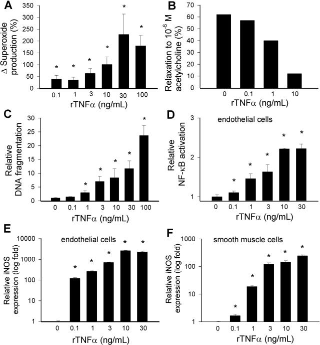
Figure 6.
Concentration dependence of the vascular effects of TNF-α. A and B: Superoxide production (A; measured by the lucigenin chemiluminescence method) and relaxations to acetylcholine (B) and in ring preparations of carotid arteries of young F344 rats maintained in vessel culture (for 24 hours) in the absence and presence of TNF-α. Data are mean ± SEM (n = 4 to 6 in each group) *P < 0.05. C: DNA fragmentation in arteries of young F344 rats maintained in vessel culture (for 24 hours) in the absence and presence of TNF-α. Data are mean ± SEM (n = 4 to 6 in each group) *P < 0.05. D: Reporter gene assay showing the effects of TNF-α on NF-κΒ reporter activity in coronary arterial endothelial cells. Endothelial cells were transiently co-transfected with NF-κΒ-driven firefly luciferase and CMV-driven Renilla luciferase constructs followed by TNF-α stimulation. Cells were then lysed and subjected to luciferase activity assay. After normalization, relative luciferase activity was obtained from four independent transfections (data are mean ± SEM, *P < 0.05 versus control). E and F: Effect of TNF-α treatment (24 hours) on the expression of iNOS in coronary arterial endothelial cells (E) and smooth muscle cells (F). Analysis of mRNA expression was performed by real-time QRT-PCR. Data are mean of four independent experiments.

