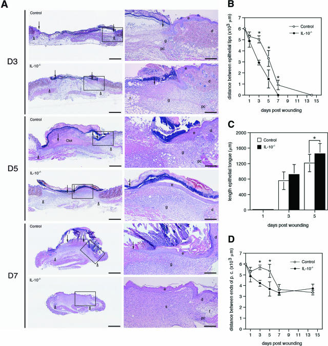
Figure 2.
IL-10 deficiency accelerates epithelialization. A: H&E staining of wounds of IL-10−/− and control mice at indicated time points after injury. In IL-10−/− mice, the length of the epithelial tongue was increased when compared with control mice. Correspondingly, the distance between the epithelial tips is reduced in knockout versus control mice. Whereas in control mice, the day 5 wound is filled with a fibrin-rich clot, the clot has been replaced by a cellular and highly vascularized granulation tissue in IL-10−/− mice; arrows point to the tips of epithelial tongues, arrowheads indicate wound edges defined by the presence of hair follicles in nonwounded skin. B: Distance between the epithelial tips (*P < 0.04); C: length of the epithelial tongue (*P < 0.018); D: distance between the edges of the panniculus carnosus (*P < 0.001) in wounds of IL-10−/− and control mice. Data are expressed as mean ± SD, n = 8 wounds for each time point and group. e, epidermis; d, dermis; f, subcutaneous fat layer; pc, panniculus carnosus; g, granulation tissue. Scale bars: 1 mm (left); 250 μm (right). Original magnifications: ×12.5 (A, left); ×50 (A, right).

