Figure 1.
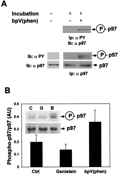
Tyrosine phosphorylation of p97. (A) LDM were incubated or not using tER assembly conditions as previously described (10–12) in the presence or absence of 100 μM bpV(phen). Solubilized membranes were immunoprecipitated either with antiphosphotyrosine antibodies (Ip: αPY, top gel) or with anti-p97 antibodies (Ip: αp97, middle and lower gels) followed by immunoblotting using anti-p97 (Ib: αp97, top and bottom gels) or anti-phosphotyrosine antibodies (Ib: αPY, middle gel) and revealed by chemiluminescence (ECL). A representative experiment is shown (n = 4) with similar results observed each time. (B) LDM were incubated in the same medium described above but containing 100 μCi [γ-32P]ATP and the absence or presence of either 100 μM bpV(phen) or 200 μM genistein. After p97 immunoprecipitation, the proteins in the immunoprecipitate were separated by SDS/PAGE and transferred onto nitrocellulose membrane. The membrane was subjected to radioautographic analysis using X-Omat AR film (top gel), then probed with anti-p97 and revealed by ECL (bottom gel). Densitometric analysis of the x-ray films was performed, and the representation is the mean of three independent experiments ± SD for the three incubation conditions.

