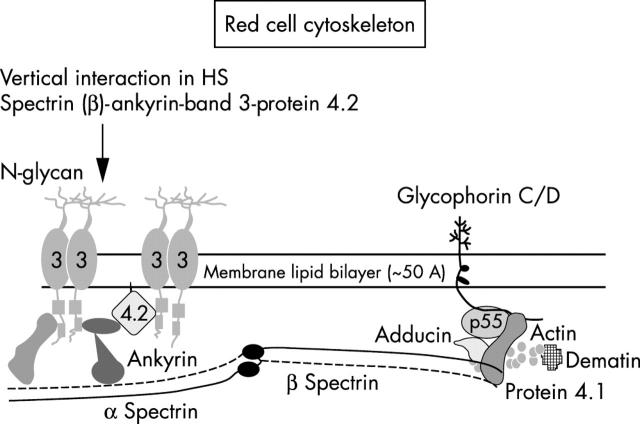
Figure 1.
Schematic presentation of the structural organisation of red cell cytoskeleton. ß Spectrin is the key component in that it pairs with α spectrin to form a heterodimer, and it has binding sites for ankyrin and protein 4.1. The common protein defects are associated with spectrin (α and/or ß), ankyrin, band 3 protein, and protein 4.1.

