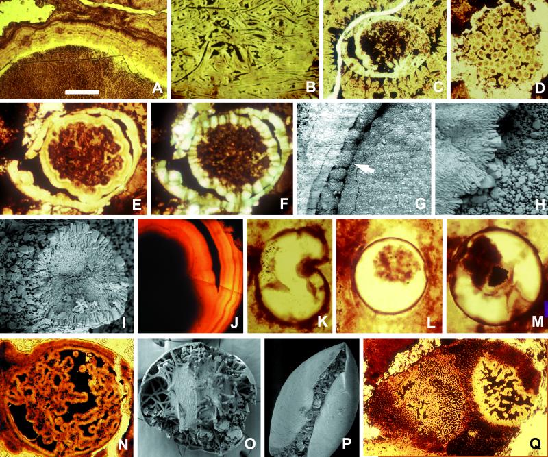
Figure 1.
Diagenetic modifications of microfossils in the Doushantuo Formation. Specimens illustrated in A–J and N–Q are from phosphorites at Weng'an, South China; K-M occur in chert nodules. Phosphatic rims occur on algal thalli (A), cyanobacterial filaments (B), the inner surfaces and collapsed contents of acanthomorphic acritarch vesicles (C), and small coccoidal cells (D). Spheroidal fossil containing diagenetically formed inner and outer rims that display crystal zonation (E) and regular orientation (F, cross-polarization); note how the inner rim is templated on the surface of preserved organic contents. (G) Phosphatic spherulitic coating on the inner surface of a vesicle (arrow), forming bulbous microstructures similar to those interpreted by Chen et al. (7) as large ectodermal cells. (H) The spot marked by an arrow in G is magnified to show crystal forms and orientation. (I) Cross section of phosphatized filament, again illustrating crystal forms and orientation. (J) Bilayered spheroidal structure similar to that in E, showing clear evidence of zoned crystal growth. (K) Silicified organic-walled vesicle with invagination produced by postmortem deformation; if phosphatized, the specimen would resemble forms interpreted as bilaterian gastrulae by Chen et al. (7); viewed in thin section, the phosphatized fossil with postmortem infolding in P would also resemble proposed gastrulae. (L and M) Individual organic-walled algal vesicles with partially collapsed internal contents, drawn from a large population of 90- to 150-μm fossils in Doushantuo cherts; postmortem phosphatization would yield structures with a size range, internal morphology, and crystal orientation comparable to those used by Chen et al. (7) to infer eumetazoan origins. (N) Internally complex spheroid, similar in organization to specimens interpreted as anthozoan planulae by Chen et al. (7), but not interpretable in biologically meaningful terms; the SEM image in O shows diagenetically phosphatized filaments and other internal structures that, viewed in thin section, would resemble N. (Q) Multicellular algal thallus (the small dark structures are cell lumens) containing a decay feature comparable in organization to structures interpreted as anthozoan planulae by Chen et al. (7). (The scale bar in A represents 100 μm for A and B; 60 μm for C, N, and Q; 80 μm for D; 50 μm for E and F; 40 μm for G, K, and L; 4 μm for H and I; 20 μm for J; 30 μm for M; 200 μm for O; and 250 μm for P.)

