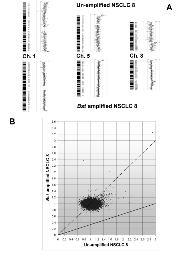
Figure 6.

Array CGH of NSCLC before and after Bst amplification. (A) Data for chromosomes 1, 5 & 8 are displayed as a karyotype diagram with values corresponding to log2 ratio of Cy5/Cy3 spot signal (SeeGH v1.6). The genome profile following Bst amplification was similar to the profile of the original sample. Clones with log2 ratio <0.5 at 1p escaped detection following Bst amplification. (B) Scatter diagram comparing ratio of Cy5/Cy3 spot signal of NSCLC 8 before and after Bst amplification. Solid line: expected 3 fold representational distortion. Dashed line: desired (1:1) ratio of ideal WGA devoid of representational distortion. Comparison of the signal ratio for NSCLC 8 before and after Bst amplification shows it is near ideal (1:1) ratio.
