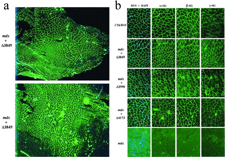
Figure 2.
IF analysis of the dystrophin and DAP complexes in gastrocnemius muscle. (a) Cryosections of mdx muscle, at 3 months after treatment with construct AAV-MCK-Δ3849 or AAV-MCK-Δ3990, were IF-stained with an antibody against dystrophin (green) and then counterstained for cell nuclei with DAPI (blue). Photos were taken with a ×4 microscope lens. Note the widespread minidystrophin expression and peripheral nucleation in a majority of the myofibers. Also note the extensive central nucleation in minidystrophin-negative areas. (b) Cryosections of muscles from 15-week-old normal C57/B10 mice, from mdx mice treated either with vector AAV-MCK-Δ3849, AAV-MCK-Δ3990, or AAV-MCK-Δ4173, or from untreated mdx mice were IF-stained with antibodies for dystrophin (green) and counterstained with DAPI (blue) for nuclei (DYS + DAPI). Note the lack of central myonuclei. The consecutive sections also were stained with antibodies for α-sarcoglycan (α-SG), β-sarcoglycan (β-SG), and γ-sarcoglycan (γ-SG). Photographs were taken with a ×20 lens.

