
Fig. 2.
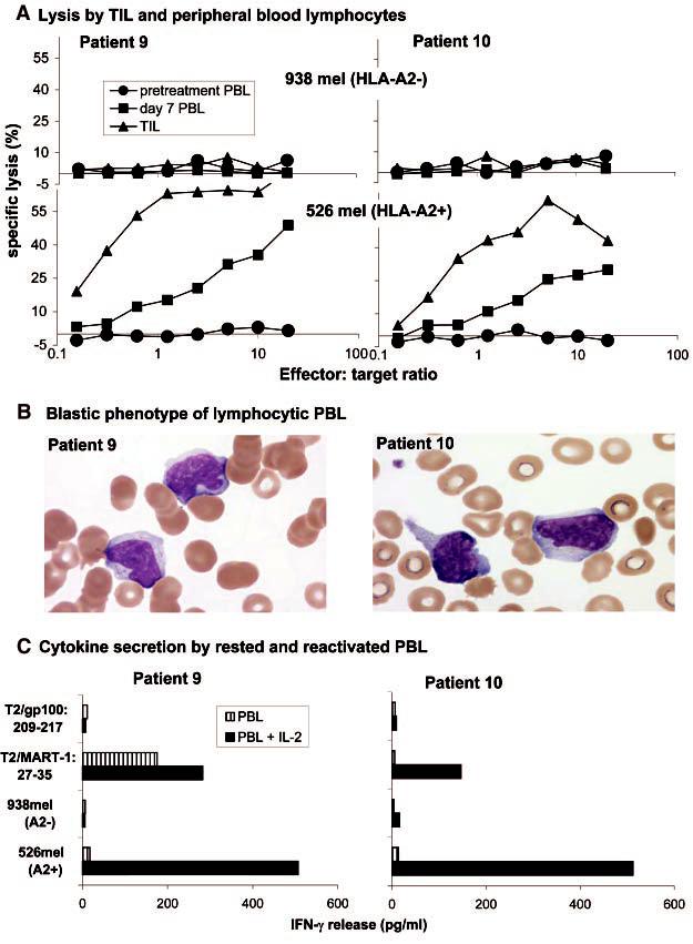
(A) The infused TILs (triangles) and PBLs collected 7 days after cell transfer (squares), but not pretreatment PBLs (circles), from patient 9 (left) and patient 10 (right) specifically lysed the HLA-A2+ melanoma cell line 526 (lowergraphs) but not the HLA-A2- melanoma cell line 938 (uppergraphs) (11). (B) Blood smears obtained from patient 9 (left) and patient 10 (right) during the lymphocytic episodes showed the highly activated phenotype of the lymphocytes, which show irregular and hyperchromatic nuclei, a high nuclear-to-cytoplasmic ratio, toxic granulation, and the presence of Dohle bodies. (C) PBLs collected 9 days (patient 9, left) or8 days (patient, 10 right) after cell transfer were cultured overnight in the absence (striped boxes) or presence (solid boxes) of 600 IU/ml of IL-2, washed extensively, and tested forcytokine release when stimulated with the indicated target cells.